ಚಿತ್ರ:Homeostasis del eritrocito y la hemoglobina.png
ಈ ಮುನ್ನೋಟ ಗಾತ್ರ:೪೫೦ × ೬೦೦ ಪಿಕ್ಸೆಲ್ಗಳು. ಇತರ ರೆಸಲ್ಯೂಶನ್ಗಳು: ೧೮೦ × ೨೪೦ ಪಿಕ್ಸೆಲ್ಗಳು | ೩೬೦ × ೪೮೦ ಪಿಕ್ಸೆಲ್ಗಳು | ೫೭೬ × ೭೬೮ ಪಿಕ್ಸೆಲ್ಗಳು | ೭೬೮ × ೧,೦೨೪ ಪಿಕ್ಸೆಲ್ಗಳು | ೧,೩೬೧ × ೧,೮೧೪ ಪಿಕ್ಸೆಲ್ಗಳು.
ಮೂಲ ಕಡತ (೧,೩೬೧ × ೧,೮೧೪ ಚಿತ್ರಬಿಂದು, ಫೈಲಿನ ಗಾತ್ರ: ೯೧೮ KB, MIME ಪ್ರಕಾರ: image/png)
ಈ ಫೈಲು ವಿಕಿಮೀಡಿಯ ಕಾಮನ್ಸ್ನಲ್ಲಿ ಇರುವುದು. ಅಲ್ಲಿನ ವಿವರಣೆ ಪುಟವನ್ನೇ ಕೆಳಗೆ ತೋರಿಸಲಾಗಿದೆ. ಕಾಮನ್ಸ್ ಕೃತಿಸ್ವಾಮ್ಯತೆಯಿಂದ ಮುಕ್ತ ಫೈಲುಗಳ ಒಂದು ಆಗರ. ಅಲ್ಲಿ ನೀವೂ ಸಹಕರಿಸಬಹುದು. |
| ವಿವರ |
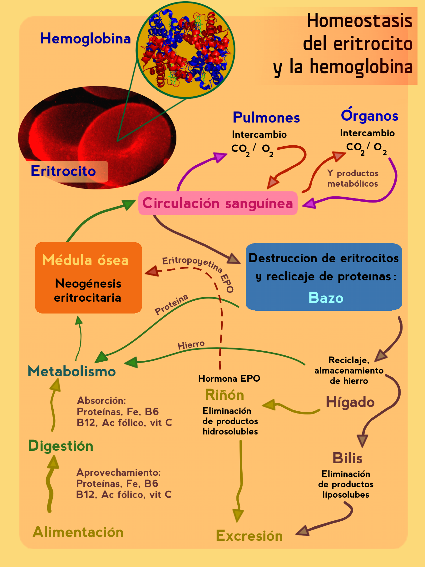
Nueva versión (en español) del gráfico FisiologiaEmoglobina.jpg de Peter Forster. en:Category:Protein images |
| ದಿನಾಂಕ | (UTC) |
| ಆಕರ | |
| ಕರ್ತೃ |
|
| This is a retouched picture, which means that it has been digitally altered from its original version. The original can be viewed here: FisiologiaEmoglobina.jpg:
|
ಈ ಕಡತ ಕ್ರಿಯೇಟಿವ್ ಕಾಮನ್ಸ್ Attribution
-Share Alike 3.0 Unported ಪರವಾನಗಿ ಹೊಂದಿದೆ.
- ನೀವು ಮುಕ್ತ:
- ಹಂಚಿಕೆಗೆ – ಕೆಲಸವನ್ನು ನಕಲು ಮಾಡಲು, ವಿತರಣೆ ಮತ್ತು ಸಾಗಿಸಲು
- ರೀಮಿಕ್ಸ್ ಮಾಡಲು – ಕೆಲಸವನ್ನು ಬಳಸಿಕೊಳ್ಳಲು
- ಈ ಕೆಳಗಿನ ಷರತ್ತುಗಳಲ್ಲಿ:
- ವೈಶಿಷ್ಟ್ಯ – ನೀವು ಸೂಕ್ತವಾದ ಕ್ರೆಡಿಟ್ ನೀಡಬೇಕು, ಪರವಾನಗಿಗೆ ಲಿಂಕ್ ಅನ್ನು ಒದಗಿಸಬೇಕು ಮತ್ತು ಯಾವುದೇ ಬದಲಾವಣೆಗಳನ್ನು ಮಾಡಿದ್ದರೆ ಸೂಚಿಸಬೇಕು. ನೀವು ಯಾವುದೇ ಸಮಂಜಸವಾದ ರೀತಿಯಲ್ಲಿ ಮಾಡಬಹುದು, ಆದರೆ ಪರವಾನಗಿದಾರರು ನಿಮ್ಮನ್ನು ಅಥವಾ ನಿಮ್ಮ ಯಾವುದೇ ಬಳಕೆಯನ್ನು ಅನುಮೋದಿಸಿದಂತೆ ರೀತಿಯಲ್ಲಿ ಉಪಯೋಗಿಸಬಾರದು.
- ಇರುವುದರಂತೆಯೇ ಹಂಚು – ನೀವು ರೀಮಿಕ್ಸ್ ಮಾಡಿದರೆ, ರೂಪಾಂತರಗೊಳಿಸಿದರೆ ಅಥವಾ ವಸ್ತುವಿನ ಮೇಲೆ ನಿರ್ಮಿಸಿದರೆ, ನಿಮ್ಮ ಕೊಡುಗೆಗಳನ್ನು ನೀವು ಮೂಲದಂತೆ ಅದೇ ಅಥವಾ ಹೊಂದಾಣಿಕೆಯ ಪರವಾನಗಿ ಅಡಿಯಲ್ಲಿ ವಿತರಿಸಬೇಕು.
Original upload log
This image is a derivative work of the following images:
- File:FisiologiaEmoglobina.jpg licensed with Cc-by-2.5
- 2006-10-29T06:01:35Z Peter Forster 526x600 (31104 Bytes) {{Information| |Description=Grafica: Medicina: Fisiologia: Ematologia: omeostasi, eritrociti, emoglobina, midollo osseo circolazione sanguigna, milza, fegato, rene, proteine, ferro Fe, eritropoietina EPO, vitamina, B6, B12, C
- 2006-10-28T05:57:48Z Peter Forster 526x600 (31104 Bytes) {{Information| |Description=Grafica: Medicina: Fisiologia: Ematologia: omeostasi, eritrociti, emoglobina, midollo osseo circolazione sanguigna, milza, fegato, rene, proteine, ferro Fe, eritropoietina EPO, vitamina, B6, B12, C
- 2006-10-19T12:22:45Z Peter Forster 526x600 (273771 Bytes) {{Information| |Description=midollo osseo, circolazione sanguina, polmoni, organi, milza, fegato, reni, bile, escrezione, alimentazione, proteine, Fe ferro, vitamina B6, vitamina B12, acido folico, vitamina C, digestione, met
- File:1GZX_Haemoglobin.png licensed with Cc-by-sa-3.0-migrated, Cc-by-sa-3.0-migrated-with-disclaimers, GFDL, GFDL-en, GFDL-self-en
- 2007-06-24T20:54:46Z PatríciaR 1600x1600 (1030152 Bytes) {{Information |Description=By Richard Wheeler ([[:en:User:Zephyris|Zephyris]]) 2007. Created with [[:en:pymol]] from . [[:en:Category:Protein images]] |Source=Originally from [http://en.wikipedia.org en.wikipedia]; descript
- File:SEM_blood_cells.jpg licensed with PD-USGov
- 2006-10-07T20:27:59Z DO11.10 1800x2239 (1398351 Bytes)
- 2006-10-04T03:00:29Z DO11.10 1800x2239 (1012422 Bytes) {{Information |Description=This is a scanning electron microscope image from normal circulating human blood. One can see red blood cells, several white blood cells including lymphocytes, a monocyte, a neutrophil, and many sma
- 2006-10-04T01:09:54Z DO11.10 500x326 (36614 Bytes) {{Information |Description= A three-dimensional ultrastructural image analysis of a T-lymphocyte (right), a platelet (center) and a red blood cell (left), using a Hitachi S-570 scanning electron microscope (SEM) equipped with
Uploaded with derivativeFX
ಕಡತದ ಇತಿಹಾಸ
ದಿನ/ಕಾಲ ಒತ್ತಿದರೆ ಆ ಸಮಯದಲ್ಲಿ ಈ ಕಡತದ ವಸ್ತುಸ್ಥಿತಿ ತೋರುತ್ತದೆ.
| ದಿನ/ಕಾಲ | ಕಿರುನೋಟ | ಆಯಾಮಗಳು | ಬಳಕೆದಾರ | ಟಿಪ್ಪಣಿ | |
|---|---|---|---|---|---|
| ಪ್ರಸಕ್ತ | ೦೭:೨೭, ೬ ಫೆಬ್ರವರಿ ೨೦೧೦ | | ೧,೩೬೧ × ೧,೮೧೪ (೯೧೮ KB) | Neotex555 | cambioa version en PNG |
| ೦೭:೧೫, ೬ ಫೆಬ್ರವರಿ ೨೦೧೦ |  | ೧,೩೬೧ × ೧,೮೧೪ (೩೬೭ KB) | Neotex555 | {{Information |Description=Nueva versión del gráfico FisiologiaEmoglobina.jpg de Peter Forster. Pedido por [http://es.wikipedia.org/wiki/Usuario:Rjgalindo] en:Category:Protein images This is a scanning electron microscope image from normal circulat |
ಕಡತ ಬಳಕೆ
ಈ ಕೆಳಗಿನ ಪುಟವು ಈ ಚಿತ್ರಕ್ಕೆ ಸಂಪರ್ಕ ಹೊಂದಿದೆ:
ಜಾಗತಿಕ ಕಡತ ಉಪಯೋಗ
ಈ ಕಡತವನ್ನು ಕೆಳಗಿನ ಬೇರೆ ವಿಕಿಗಳೂ ಉಪಯೋಗಿಸುತ್ತಿವೆ:
- ca.wikipedia.org ಮೇಲೆ ಬಳಕೆ
- es.wikipedia.org ಮೇಲೆ ಬಳಕೆ